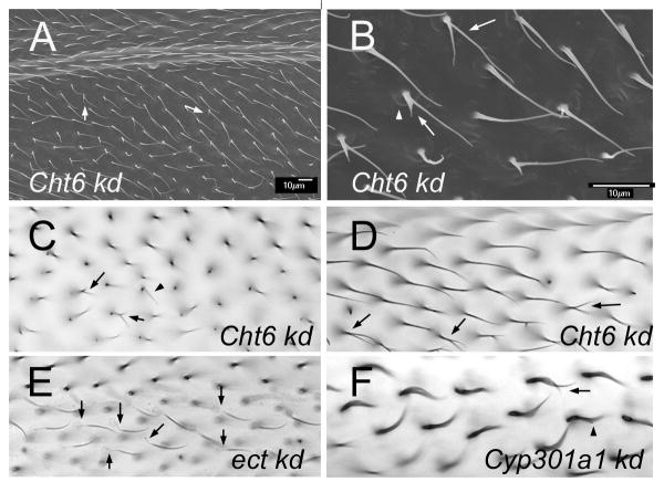
Fig. 7.
(A) An SEM of an adult ptc-Gal4 UAS-Cht6 RNAi wing. Arrows point to branched hairs. Note the poor alignment of neighboring hairs. (B). A higher magnification image of a region of A. The arro points to a split hair. The arrowhead points to a “cup” at the base of the hair. (C). A brightfield micrograph of an adult ptc-Gal4 UAS-Cht6 RNAi wing. The arrows point to branched and thin hairs. (D). A brightfield micrograph of an adult ptc-Gal4 UAS-Cht6 RNAi wing. The arrows point to branched hairs. (E). A brightfield micrograph of an adult ptc-Gal4 UAS-ect RNAi wing. The arrows point to hairs that show the “ect” phenotype of hairs with a faint and wimpy proximal region. (F). A brightfield micrograph of an adult ptc-Gal4 UAS-Cyp301a RNAi wing. The arrows point to split hairs. Note the curved shape of all hairs in this wing region.

