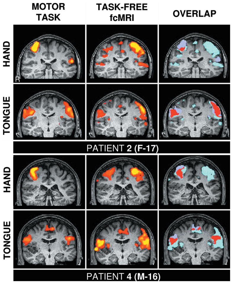
Fig. 2.

Functional mapping based on fcMR imaging is anatomically specific. These studies are comparisons of hand and tongue motor regions defined by actual motor task movements (left columns) and task-free fcMR imaging (center columns). The overlap of the two techniques is shown in red (right columns). The upper panels show data from the patient in Case 2, and the lower panels from the patient in Case 4. The right hemisphere is displayed on the right side of the panel. Note the systematic shift of the location of the hand and tongue regions in each patient, which is present for the fcMR analysis. The fcMR analysis of the hand region in the patient in Case 2 is less stable than other measures, possibly due to the location of the seizure activity (see text).
