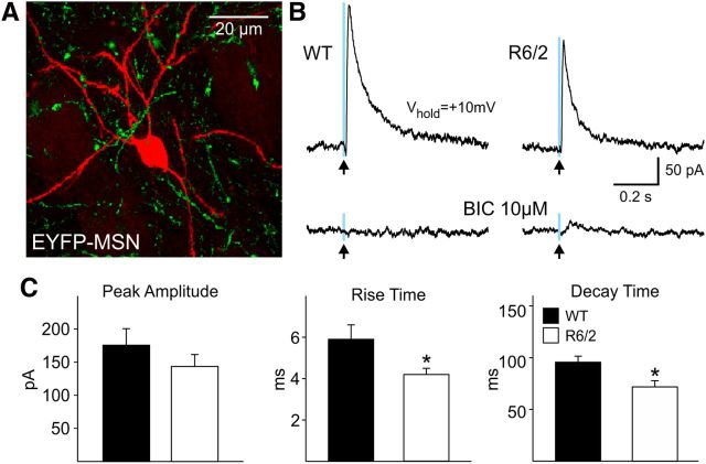
Figure 9.
A. Biocytin-filled MSN (red) and the EYFP expression (green) in axons from SOM interneurons. B, Outward currents evoked in an MSN from a WT and an R6/2 mouse in response to blue light stimulation (0.5 ms duration, 8 mW power) at a holding potential of +10 mV. These currents were blocked by BIC (10 μm) and were not evoked by yellow light (data not shown). C, Bar graphs show that mean peak amplitudes were similar but there were faster rise (p < 0.05) and decay times (p < 0.05) in MSNs from R6/2 compared with those from WT mice.

