
Figure 3.
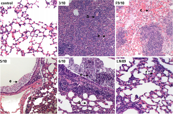
Comparison of Lung pathology in mice infected with different H1N1/2009 viruses on 5 dpi. In 3/10-infected mice lungs, signs of acute interstitial pneumonia with infiltration of large amount of lymphocytes (arrow a) and atelectasis (arrow b) were present. Lung tissues in lesion areas of F9/10-infected mice show acute pneumonia with alveolar wall thickening, large amounts of infiltration of inflammatory cells and bleeding (arrow c). The pathological changes of lung tissues infected with 5/10 and 6/10 viruses just show certain alveolar wall thickening (arrow d) and bronchial exudates (arrow e). LN/09-infected mice only show slight alveolar wall thickening in lung lesions (arrow f). Images were obtained on an Olympus BX-50 light microscope at 50-fold original magnifications (50×).