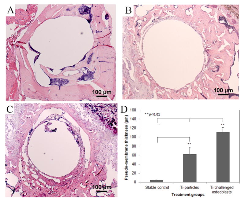
Fig. 5.

(A–C) represent the histological appearances of pin-implanted tibiae at 4 weeks following different treatments (40×): (A) stable pin implantation without particles stimulation; (B) Ti particles treated prosthesis failure control; (C) the injection of Ti particles-challenged OBs significantly result in more severe inflammation with thicker membrane generation. (D) The comparison of periprosthetic membrane thickness in response to different treatments (**p<0.01).
