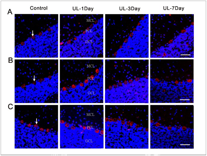
Figure 7. Localization and distribution of histamine receptors in the flocculus.
Representative confocal images showing the expression and localization of the H1 (A), H2 (B) and H3 (C) receptors in the same area of the ipsi-lesional flocculus following UL or sham operation at different post-operative days. Arrows show the H1-, H2- and H3 receptors -positive neurons. UL-1day, 1 day after UL; UL-3day, 3 day after UL; UL-7day, 7 day after UL; Control, with intact labyrinths; GCL, granule cell layer; PCL, Purkinje cell layer; MCL, molecular cell layer. Calibration bar = 40 µm.

