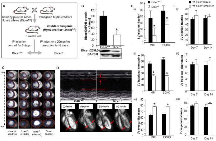
Figure 1. Cardiac specific Dicer deletion leads to cardiac hypertrophy and impaired cardiac function.
(A) Generation of Myh6-cre/Esr1-Dicerfl/fl mice. (B) Western Blot showing reduced Dicer expression following tamoxifen injection. (C) 11.7T images of short axis at diastole and systole of 1.0 mm slices stacked along columns from base to apex of the left ventricle of both Dicer+/+ and Dicer−/− mice. Blue contour lines indicate LV volume. (D) Representative M-mode images of Dicer+/+ and Dicer−/− mice. The data indicates decreased contractility and increased left ventricular (LV) internal dimensions in Dicer−/− mice. 11.7T MRI images of long axis at diastole (i & iii), systole (ii & iv) cardiac 11.7T MRI images in Dicer+/+ and Dicer−/− mice. The diastole of Dicer−/− mice was enlarged due to insufficient contraction of the heart (shown by arrows). (E) Quantification of LV fractional shortening, LV myocardial mass, and LV ejection fraction (EF) by MRI and ECHO. Solid bars represent Dicer+/+ and open bars represent Dicer−/−. (F) (i) Ejection fraction, (ii) fractional shortening and (iii) LV mass in wild type corn oil or tamoxifen injected mice.

