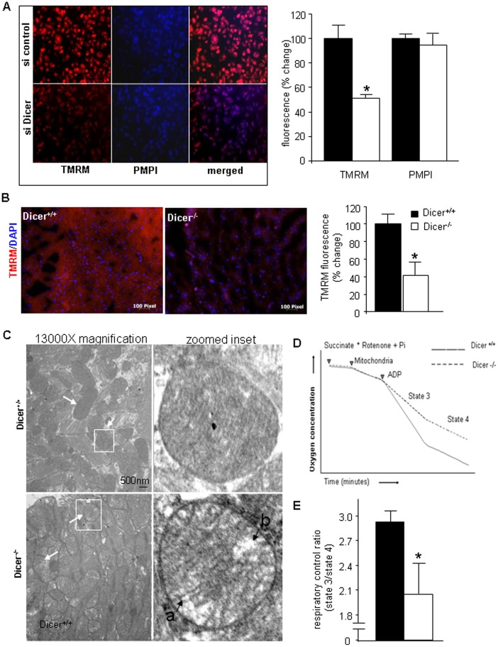
Figure 3. Dicer deletion in the adult heart leads to mitochondrial dysfunction.
(A) TMRM assay in HL-1 cells transfected with si-control and si-Dicer. (B) TMRM assay on Dicer+/+ and Dicer−/− heart sections. Bar graph quantifying TMRM fluorescence demonstrates lower TMRM fluorescence in Dicer−/− hearts (n = 3). (C) Representative TEM images of mitochondria from heart tissue. Arrows indicate mitochondria. Single mitochondria in white boxes have been zoomed into in the right panel. Labels a) indicate increased inter-cristae space as compared to the mitochondria in the Dicer+/+ heart and b) points to loss of integrity of the cristae structure. (D) Representative diagram showing experiment for RCR measurements from isolated mitochondria. (E) Respiratory coupling ratio (RCR) was significantly lower in Dicer−/− mice indicating mitochondrial dysfunction.

