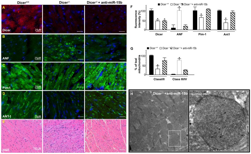
Figure 8. Suppression of induced miRNA-15b attenuates loss of Pim-1 and improves mitochondrial integrity.
Immunohistochemical comparison of (A) Dicer (B) ANF, (C) Pim-1(D) ANT-1 (E) Comparison of cardiac histopathology sections stained with hematoxylin and eosin (H&E) (F) Bar graph showing quantification of IHC images. Image analysis software (Axiovision 4.3, Zeiss, Germany) was used to quantify fluorescence intensity (fluorescent pixels) and analyzed as a percent change in relative fluorescence unit (RFU). (n = 3) (G) Comparison between % of total mitochondria belonging to Class I/II or Class III/IV. (H) Representative TEM images of mitochondria from Dicer depleted heart tissue treated with anti-miR-15b. Bar graphs represent equal SD on both sides of the mean.

