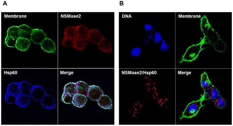
Figure 8. Localization of N-SMase2, Hsp60 and N-SMase2/Hsp60 complex.
HEK-293 cells were seeded and transfected with N-SMase2 in 35-mm confocal dishes. After 24 h, (A) Cells were fixed and stained with wheat germ agglutinin (green), anti-N-SMase2 (red) and anti-HSP60 (blue) as described under ‘Materials and Methods’. (B) On the other hand, the cells were stained with wheat germ agglutinin (green). To visualize a complex of Hsp60 and N-SMase2 as heterodimers we performed an In situ proximity ligation assay using proximity probes against Hsp60 and N-SMase2. The cells were stained with DAPI (blue) to visualize the nucleus.

