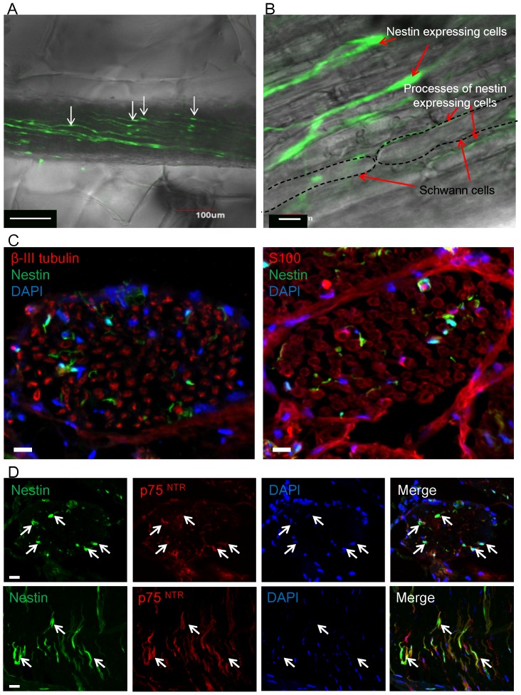
Figure 1. Location and characteristics of ND-GFP-expressing cells in the sciatic nerve.
(A) The first day of Gelfoam® histoculture of a sciatic nerve bundle removed from the ND-GFP transgenic mouse. The sciatic nerve contained ND-GFP-expressing cells which had long processes (white arrows). Bar: 100 µm. (B) High-magnification image of the sciatic nerve showed that ND-GFP expressing cells and their processes are located between Schwann cells. Bar: 10 µm. (C) Transverse sections of the sciatic nerve of the ND-GFP mouse. The ND-GFP-expressing cells did not express β-III tubulin and S100. Bar: 10 µm. (D) Transverse and longitudinal sections of the sciatic nerve of the ND-GFP mouse. The ND-GFP-expressing cells (white arrows) co-expressed p75NTR. Bar: 10 µm.

