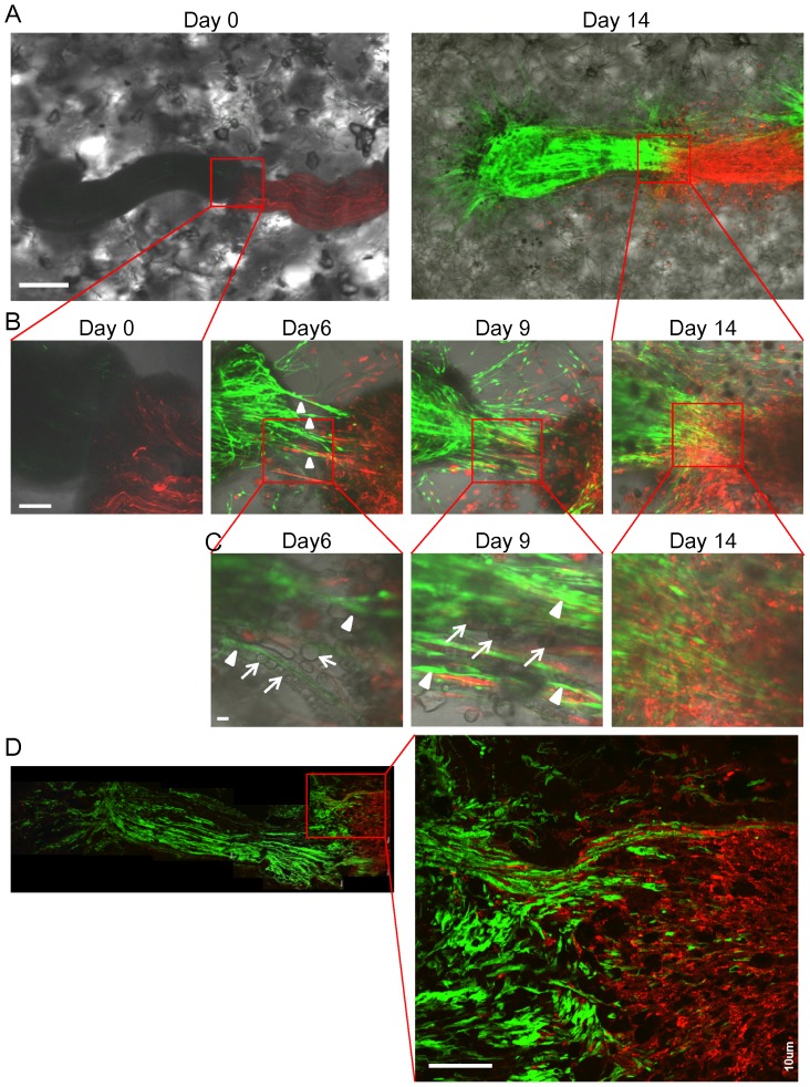
Figure 6. Intermingling of growing sciatic nerve in 3D Gelfoam® histoculture.
(A) A sciatic nerve from a ND-GFP transgenic mouse was placed on Gelfoam® next to the sciatic nerve from an RFP transgenic mouse. At day 14, the sciatic nerve from the ND-GFP mouse was enriched with ND-GFP-expressing cells and intermingled with to the RFP-expressing sciatic nerve. Bar: 500 µm. (B) Magnified images of the area inside the box in Figure 6A show that the ND-GFP-expressing cells proliferated in fibers growing from the nerve extending toward the other sciatic nerve. At day 9, the thickest fibers appeared between both sciatic nerves. At day 14, the two nerves intermingling with each other. Bar: 100 µm. (C) Magnified images of the area inside the box in Figure 6B show that the fibers consisted of ND-GFP-expressing spindle cells (white arrow heads) and ND-GFP-negative spherical cells (white arrows). The spherical cells formed a line between both sciatic nerves and ND-GFP-expressing spindle cells extended among the lines. Bar: 10 µm. (D) A section of the intermingling two nerves. High-magnification images show that ND-GFP-expressing fibers growing from the sciatic nerve from the ND-GFP mouse invaded deeply into the RFP-expressing sciatic nerve. Bar: 100 µm.

