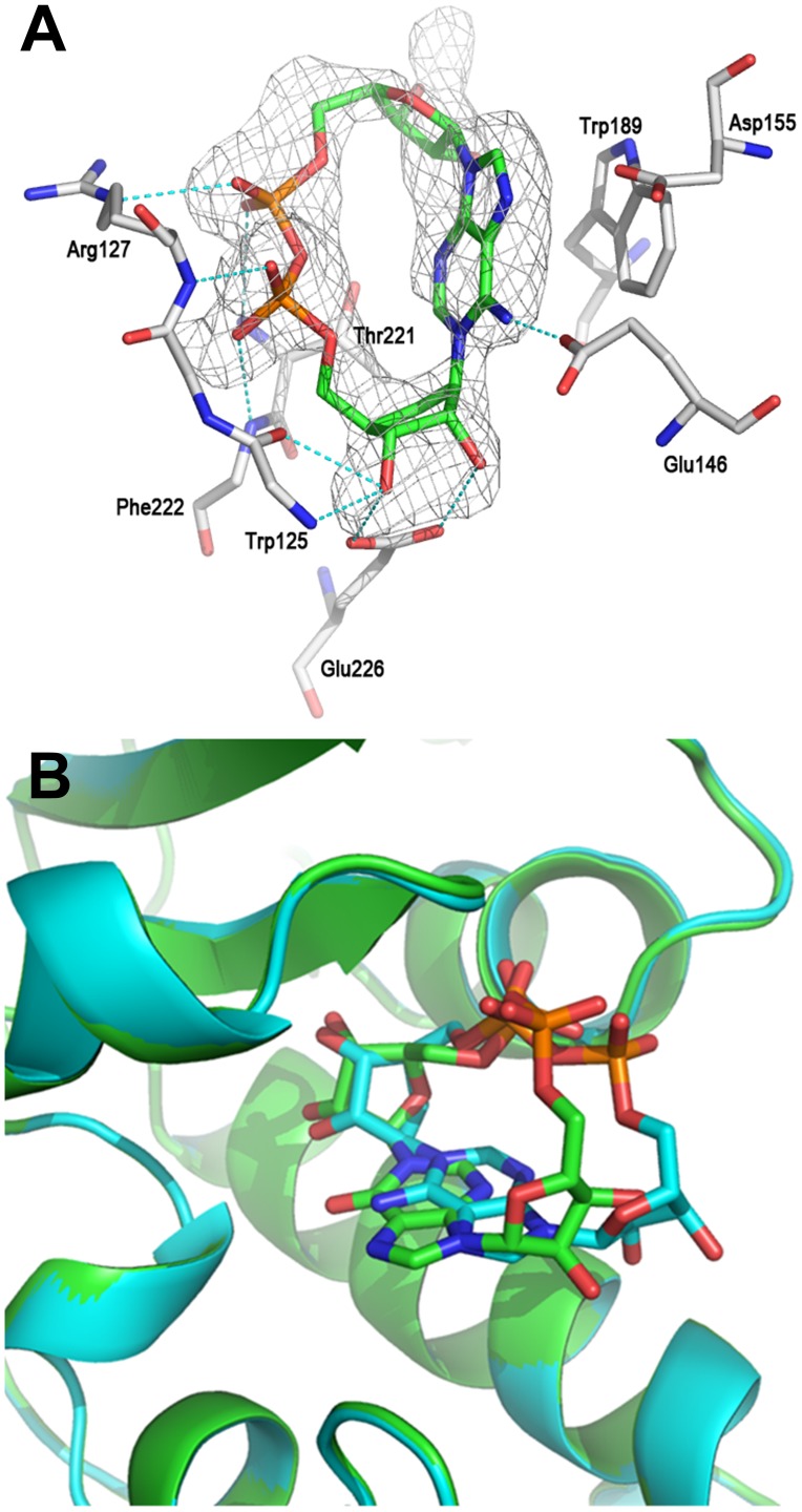
Figure 8. The crystal structure of CD38 with cADPcR.
(A) Structure of cADPcR complexed with wild-type CD38. The σA weighted Fo-Fc different electron density is shown as gray isomesh contoured at 2.5σ. H bonds are shown as dashed lines and colored in cyan (B) Binding comparison between N1-cIDPR (carbons in green) and cADPcR (carbons in cyan) complexed with CD38, showing overlap of their “northern” ribosyl phosphate motifs.

