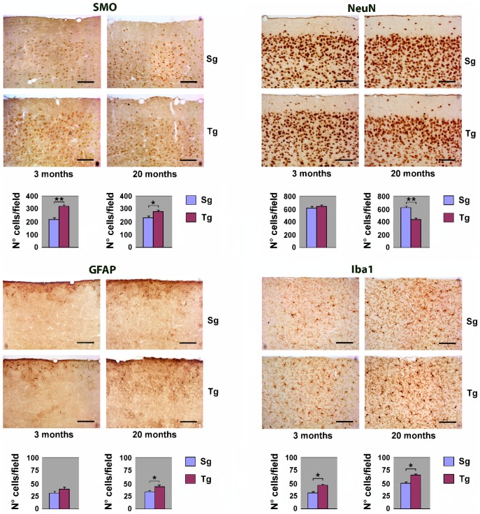
Figure 3. Immunohystochemical analysis of neocortex from Tg and Sg mice.
Sagittal brain slices from JoSMOrec mice were stained with antibodies directed against SMO, NeuN, GFAP and Iba1. Slides of neocortex from 3 and 20 months old mice were analyzed. Cell counting is expressed as number of positive cells per 0.24 mm2 area. The p values were measured with the one-way ANOVA test and post-hoc test Bonferroni (*, p<0.05; **, p<0.01; ***, p<0.001). Sg, syngenic mice; Tg, transgenic mice.

