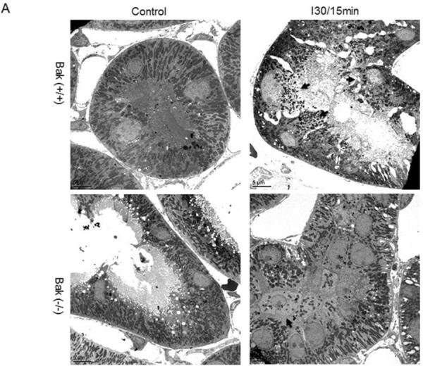
Fig. 9. Inhibition of mitochondrial fragmentation during ischemia/reperfusion in germline Bak-KO mice.


Kidney tissues were collected after 30 minutes of ischemia and 15 minutes of reperfusion and then processed for electron microscopy. (A) Representative images showing the morphology of mitochondria. Arrows point to the cells with fragmented mitochondria. (B) Quantification of the cells with mitochondrial fragmentation. The cells with filamentous and fragmented mitochondria were counted to determine the percentage of cells with mitochondrial fragmentation.
