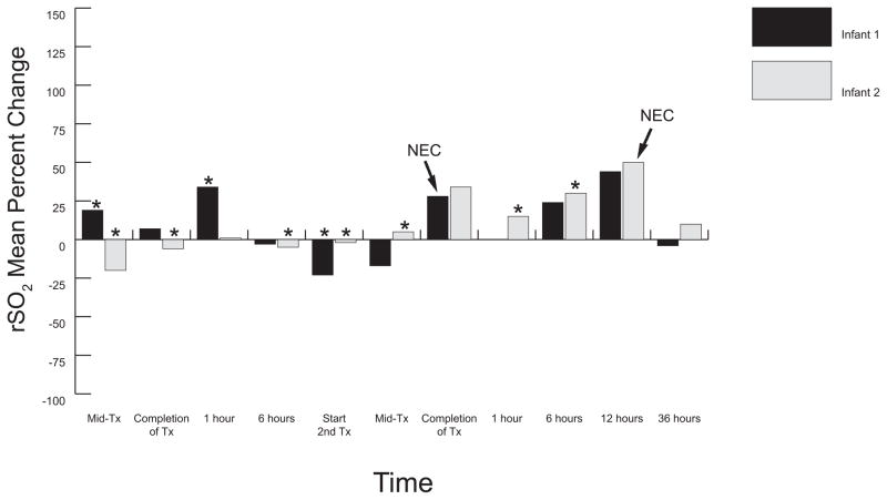
Figure 1.
Mesenteric Mean Percentage Change from Baseline: Medical TR-NEC Infants. This graph illustrates the wide mesenteric oxygenation fluctuations above and below baseline measurements during and subsequent to each transfusion event and further reveals decreased oxygenation immediately prior to TR-NEC onset and subsequent increased patterns at the time of TR-NEC onset. Infant 1 received two half-volume PRBC transfusions (7.5ml/kg each) separated by 12 hours. Infant 2 received two full volume (15ml/kg) PRBC transfusions separated by 21 hours. Infant 1 had NIRS monitor removed during resuscitation and transfer to NICU (time point 1 hour after 2nd transfusion). Tx, transfusion; TR-NEC, transfusion-related necrotizing enterocolitis; Mid-Tx, time at which 50% of total volume had infused; * enteral feeding given during specified time frame; 0 = baseline; NEC, onset of TR-NEC.

