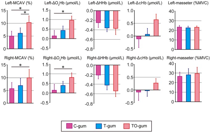
Figure 4. Summaries of hemodynamic changes in the left (top panels) and right (bottom panels) cerebral hemispheres and masseter EMG activity associated with chewing of three different types of gum.
In the hemodynamic changes, MCAV, ΔO2Hb, ΔHHb, and ΔcHb are arranged from the left to the right. Individual data (mean ± SEM) are represented as change rates of the responses between gum-chewing tests (5 min) and before the test. Statistical comparisons were performed among the three gums. *P<0.05, **P<0.01.

