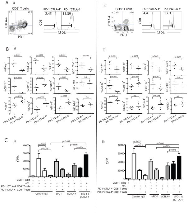
Figure 2. Co-expression of PD-1 and CTLA-4 correlates with more severe dysfunction of CT26 and ID8-VEGF antigen-specific CD8+ T cells.
A) PD-1+CTLA-4+ and PD-1+CTLA-4− CD8+ TIL populations from either CT26 (i) or ID8-VEGF (ii) tumors were sorted, labeled with CFSE, and stimulated with AH-1 or FR peptide, respectively for 3 days in the presence of irradiated splenic CD45.1− APCs. CD8+ T cell proliferation was determined by dilution of CFSE; numbers indicate the percentage of CFSElo (AH-1 or FR reactive cells). B) Frequency of CT26 (i) or ID8-VEGF (ii)-specific PD-1+CTLA-4− and PD1+CTLA-4+ CD8+ T cells showing cytokine and degranulation after peptide stimulation (n=6). PD-1+CTLA-4− and PD1+CTLA-4+ CD8+ TILs cells were further analyzed for differential phenotype and expression of other inhibitory receptors. C) PD-1+CTLA-4+ or PD-1+CTLA-4− CD8+ TILs from CT26 (i) or ID8-VEGF (ii) were stimulated with AH-1 or FR peptide, respectively and cultured with blocking antibodies for 3 days. Proliferation was determined by 3[H]-thymidine incorporation. Statistical significance was determined by Student’s t-test.

