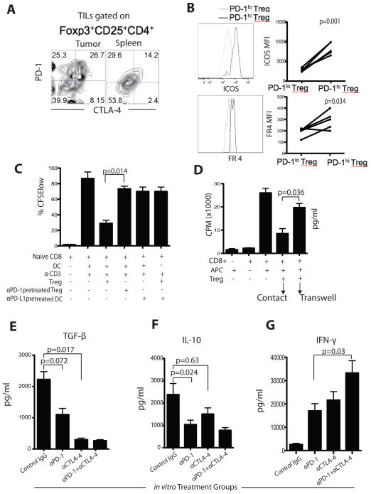
Figure 7. PD-1 and CTLA-4 pathway blockade reduces Treg-mediated suppression of CD8+ T cells in vitro and secretion of regulatory cytokines TGF-β and IL-10.
A) Representative expression of PD-1 and CTLA-4 by intra-tumoral and splenic Tregs in the same mouse. B) ICOS and folate receptor 4 (FR4) expression and MFI in PD-1hi and PD-1lo Tregs. C) TIL Tregs (± αPD-1) were co-cultured with CFSE-labeled CD8+ T cells and DCs (pretreated) in the presence of αCD3. Dividing cells were quantified after 4-days of culture by gating on the CFSElo population. D) [3H]-thymidine incorporation into effector T cells in the presence or absence of TIL Tregs. Tregs were either in direct contact with the effector cells or were separated by a transwell membrane. The whole tumor leucocytes were isolated and cultured ex vivo for 72 hours in the presence of AH-1 peptide and PD-1 and CTLA-4 blocking antibodies as indicated. After culture, supernatants were analyzed for secretion of TGF-β (E), IL-10 (F), and IFN-γ (G). Results are from 3 representative experiments. Bar graphs show mean ± SD.

