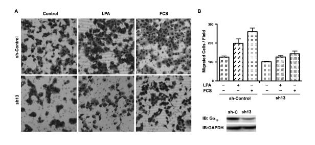
Figure 8. Effect of Silencing Gα13 on LPA- and Serum-stimulated Invasive Migration of Panc-1 Cells.
(A) Panc-1 cells in which the expression of Gα13 was silenced by shRNA to Gα13 (sh13) or cells expressing control shRNA (sh-Control) were monitored for their invasive migratory response to LPA (20 μM), 10% FCS, or serum-free medium (control) on type-1 collagen treated transwell system as described under Materials and Methods. At 20 hrs following LPA stimulation, images were obtained of random fields of view at 100X magnification. The images shown are representative of three independent experiments, each performed with triplicate fields of view. (B) Cell migration profiles were quantified by enumerating the migrated cells in a minimum of three fields. Results are presented as the number of migrated cells per field and the bars represent the mean ± SEM from three independent experiments. (C) Silencing of endogenous Gα13 was monitored by immunoblot analysis using antibodies to Gα13, probing the lysates from cells expressing control non-specific shRNA (sh-C) and cells in which the expression of Gα13-was silenced using Gα13-specific shRNA (sh13). The blot was stripped and re-probed with antibodies to GAPDH to monitor equal loading of protein.

