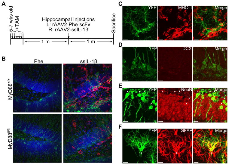
Fig. 4.
Alteration of cell fate by rAAV2-ssIL-1β induced inflammation. Experimental design showing tamoxifen (TAM) administration as well as intrahippocampal injections (A). MHC-II (red) staining demonstrating unilateral nature of inflammation in addition to YFP+ (green) cells in hippocampi that received ssIL-1β (B,C). Co-labeling of YFP+ cells with DCX to label neuroblasts (D), NeuN to label neurons (E), and GFAP to label reactive astrocytes (F). Arrowheads indicate double positive cells. Arrow indicates a NeuN−YFP+ cell. Scale bars = 10 μm.

