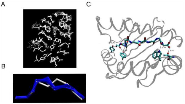
Figure 1.

Test set comparison. A. Superimposed 18 binding pockets of the MHC class molecule HLA-A*0201, obtained by X-ray crystallography. Each pocket represents the bound conformation to each of 18 peptides. The receptor residues within 5 Å from the bound peptide are considered only. B. Superimposed conformations of the 18 epitope backbones restricted by HLA-A*0201, obtained by X-ray crystallography. The peptide 2V2X is represented by white and the other 17 peptides are represented by blue. C. Four conserved hydrogen bonds between the peptide backbone and the receptor side chain.
