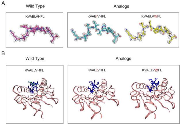
Figure 5.
The x-ray structures of the pMHC in the blind test set. A. The electron density around the wild peptide (KVAELVHFL) and two mutants (KVAEIVHFL and KVAELVWFL), bound to HLA-A*0201. The asymmetric unit of the crystals contains two copies of pMHC complexes. The electron density for the first copy is shown here and the electron density for the peptide in the second copy is shown in Figure 6. B. Crystallographic models of the three peptides bound to HLA-A*0201. The overlay of the two configurations for the wild peptide is in blue and cyan.

