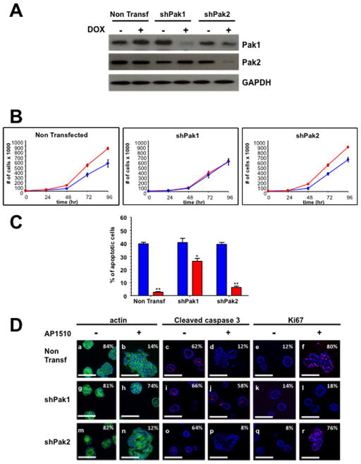
Figure 1. Pak1, but not Pak2, is required for ErbB2-mediated transformation of MCF-10A cells.
(A) Western analysis shows specific loss of Pak1 and Pak2 in shRNA infected cells. (B) Proliferation of Pak1 and Pak2 10A.ErbB2 deficient cells upon ErbB2 stimulation. Cells were seeded, harvested and counted at 0, 24, 48, 72 and 96 h. The data are representative of three independent experiments. Points, mean; bars, SD. (C) Apoptosis of Pak1 and Pak2 10A.ErbB2 deficient cells. Apoptosis was measured calculating the percent of positive Annexin V-PE cells by flow cytometry. The data are representative of three independent experiments. Bars, SD. (D) Pak1 and Pak2 in shRNA-infected 10A.ErbB2 cells were plated atop reconstituted basement membrane. Cells treated with doxycycline and stimulated with vehicle or 1 μM AP1510 on day 3 and fixed on day 12 were stained with Oregon green-phalloidin, Ki-67, or anti-cleaved caspase-3. Percentage of unilamellar acini, Ki-67-positive, and anti-cleaved caspase-3-positive acini were scored based on assessment of 50 to 60 acini per well. Bar, 50 μm.

