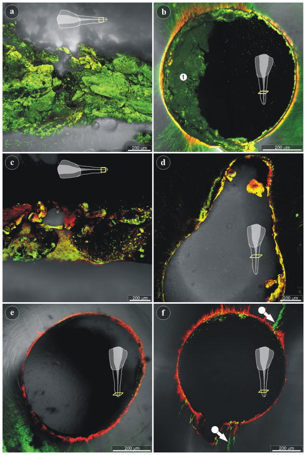
Figure 4.
cLSM imaging of untreated and treated root canals: (a) Longitudinal-section through the apical part of a root canal, as indicated in the scheme. Live/Dead® staining of an untreated specimen revealing vital biofilm (green signal). (b) The cross-section through a root canal of an untreated tooth also shows vital biofilm (area 1). (c) The longitudinal-section through the apical part of a plasma-treated root canal reveals areas with live (green) and dead (red) bacteria, while large regions have no signal and remain black. (d) Cross-section through the middle part of a plasma-treated root canal, showing little biofilm with mostly compromised bacteria (yellow signal). (e, f) The apical regions of two NaOCl-treated root canals demonstrate the almost complete removal of biofilm from the root canals walls. Only few small side-canals show persisting biofilm with viable bacteria (f).

