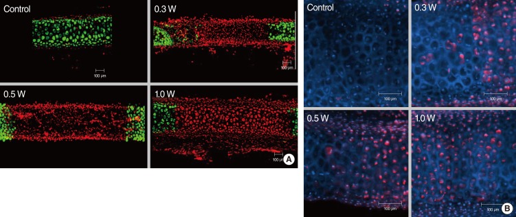
Fig. 4.
(A) Confocal images of live/dead assay of the cartilage injury after laser irradiation in rabbit auricular cartilage. Cartilage was irradiated with laser power of 0.3 W, 0.5 W, and 1.0 W for 5 seconds. The extent of damaged area increased with increase in laser power (0.3-1.0 W). (B) Hoechst & PI staining imaging of rabbit auricular cartilage following laser irradiation (laser power, 0.3-1.0 W; exposure time, 5 seconds). Blue and red stained cell indicates live and necrotic cell, respectively. This image shows that thermal injury of chondrocytes resulted in necrosis of chondrocytes rather than apoptosis.

