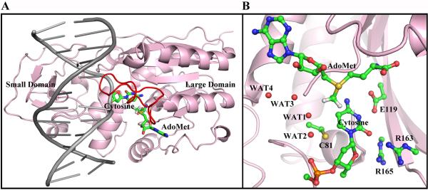
Figure 1.

(A) Ternary structure of HhaI methyltransferase (PDB ID38: 6MHT). The flipped out cytosine and the cofactor AdoMet are colored by atom. The protein is pink. The large and small domains are indicated. The DNA is gray and the catalytic loop is red. (B) The active site, including the target cytosine, catalytic Cys81 from the catalytic loop, Glu119, Arg163 and Arg165 from the large domain and the cofactor AdoMet are shown, and crystal waters are indicated. The active site structure was remodeled from the crystal structure as described in Methods.
