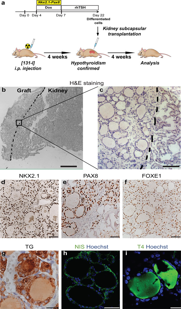
Figure 3. Grafting of mESC-derived thyroid follicles in mice.
a, Schematic diagram of protocol for mESC-derived thyroid follicles transplantation in the renal capsule of mice with radio-ablated thyroid (hypothyroid mice). b–i, Histological analysis of kidneys sections 4 weeks after grafting. Hematoxylin and eosin staining (H&E) on OCT-embedded grafted kidney showed: - localization of transplanted tissue in the cortical area of the host organ (left side) (b) - and single cuboidal epithelium organization of transplanted tissue (c); Immunohistochemistry of NKX2.1 (d), PAX8 (e), FOXE1 (f), TG (g), and immunofluorescence of NIS (h) and T4 (i) in grafted tissue. Scale bars 300 µm (b), 100 µm (c), 50 µm (d,e,f,h) and 20 µm (g,i).

