
Figure 10.
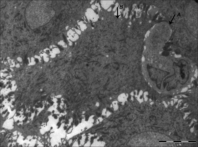
Electron micrograph of erosive OLP showing langerhans cell (A) in close proximity to keratinocyte (B) with loss of nucleus and abundant accumulation of tonofilaments – ×880
Official websites use .gov
A
.gov website belongs to an official
government organization in the United States.
Secure .gov websites use HTTPS
A lock (
) or https:// means you've safely
connected to the .gov website. Share sensitive
information only on official, secure websites.
Electron micrograph of erosive OLP showing langerhans cell (A) in close proximity to keratinocyte (B) with loss of nucleus and abundant accumulation of tonofilaments – ×880