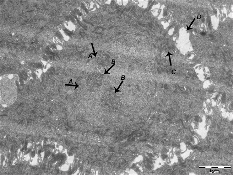
Figure 3.

Electron micrograph of nonerosive OLP showing nuclear membrane infolding (A), prominent nucleoli (B), tonofilament accumulation in the cytoplasm of the cells of spinous layer (C). Widened intercellular spaces between the cells of spinous layer (D) – ×1600
