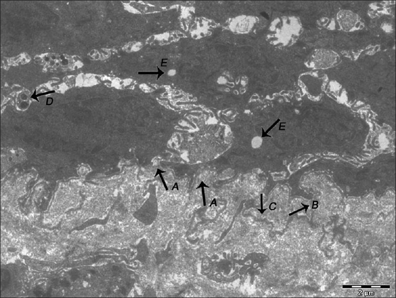
Figure 6.

Electron micrograph of erosive OLP showing basal lamina exhibiting breaks (A), duplication (B), loss of hemidesmosomes and separation of basal lamina from the surface of the basal epithelial cells (C). Keratinocytes undergoing apoptosis showing the presence of membrane-bound apoptotic bodies (D). Vacuolization is evident in the cytoplasm of keratinocyte (E) – ×1400
