Figure 7.

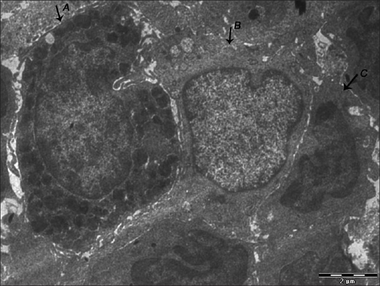
Electron micrograph of erosive OLP showing the presence of mast cell (A), langerhans cell (B), and macrophage (C) close to each other – ×1800

Electron micrograph of erosive OLP showing the presence of mast cell (A), langerhans cell (B), and macrophage (C) close to each other – ×1800