
Figure 3.
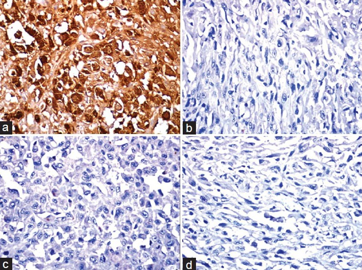
Immunohistochemical staining showing (a) Positivity of tumor cells with S-100 and negativity for (b) Pan cytokeratin, (c) HMB 45, and (d) Vimentin (×400)
Official websites use .gov
A
.gov website belongs to an official
government organization in the United States.
Secure .gov websites use HTTPS
A lock (
) or https:// means you've safely
connected to the .gov website. Share sensitive
information only on official, secure websites.
Immunohistochemical staining showing (a) Positivity of tumor cells with S-100 and negativity for (b) Pan cytokeratin, (c) HMB 45, and (d) Vimentin (×400)