Figure 2.

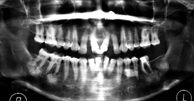
Orthopantomogram showing multiple well-defined sclerotic masses with radiolucent border in both right and left molar region of the mandible

Orthopantomogram showing multiple well-defined sclerotic masses with radiolucent border in both right and left molar region of the mandible