
Figure 2.
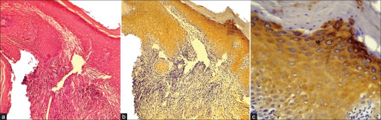
Immunohistochemical expression of Perlecan in Oral Dysplastic Epithelium (a) Oral Dysplastic Epithelium H and E ×100, (b) Immunohistochemical expression of Perlecan in suprabasal (stratum spinosum, stratum granulosum) layers IHC ×100, (c) Coalescent Immunohistochemical expression of Perlecan within the cell, at the cell surface & intercellular spaces IHC ×400. Note: Stratum corneum does not show immunoexpression of Perlecan.