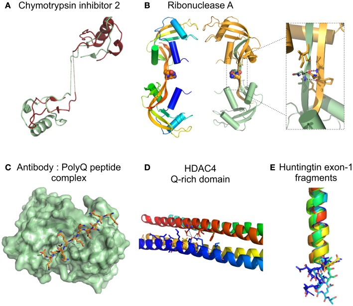
Figure 3.
Structure of proteins/protein domains containing polyQ regions. (A) Cartoon representation of the domain swapped dimer of chymotrypsin inhibitor 2 with a 4 glutamine insertion [(Chen et al., 1999); PDB accession code 1cq4], dotted lines represent the polyQ linker not visible in the X-ray crystal structure. (B) Cartoon representation of domain swapped major dimers of ribonuclease A. Inset shows a short segment resembling the polar zipper formed by asparagine residues in the linker region [(Liu et al., 2001); PDB accession code 1f0v]. (C) Surface representation Fv fragment of a monoclonal antibody in complex with a polyQ peptide shown as sticks [(Li et al., 2007a), PDB accession code 2otu]. (D) Cartoon representation of the glutamine-rich domain from HDAC4 showing details of the polar interactions (dotted lines) at the oligomer interfaces involving glutamine residues [(Guo et al., 2007), PDB accession code 2o94]. (E) Cartoon representation of the crystal structures of huntingtin exon-1 fragments observed in different crystal forms, highlighting the different orientations of the C-terminal polyQ residues shown as sticks. The 17 glutamine stretch adopts variable conformations in the structures: α helix, random coil, and extended loop. [(Kim et al., 2009), PDB accession codes 3io4, 3iow, 3iov, 3iou, 3iot, 3ior, 3io6].

