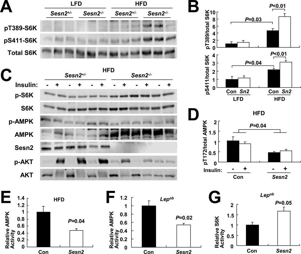
Figure 3. Sestrin2 regulates AMPK-mTORC1 signaling in liver.
(A and B) Livers from 6 months-old mice of the indicated genotypes kept on LFD or HFD for 4 months were analyzed by immunoblotting for mTORC1-dependent S6K phosphorylation at Thr389 and Ser411 (A). Ratio of phosphorylated to total S6K was quantified by densitometry and presented as bar graphs (B, n≥3). (C and D) Livers were collected from Sesn2+/− and Sesn2−/−mice kept on HFD, after 6 hrs of fasting, before (−) or 10 min after (+) insulin injection (0.8 U/kg body weight), and analyzed for protein phosphorylation and expression with indicated antibodies (C). Ratio of phosphorylated to total AMPK was presented as a bar graph (D, n=3). (E–G) AMPK (E and F) and S6K (G) activities in livers of HFD-fed Con and Sesn2−/− mice (E) or Lepob/ob/Sesn2+/− and Lepob/ob/Sesn2−/− mice (F and G) were measured by kinase assays (n=3). Levels of immunoprecipitated kinases and corresponding kinase activities are presented in Figure S3. Data are presented as means ± standard error. P values were calculated using Student’s t-test or two-way ANOVA.

