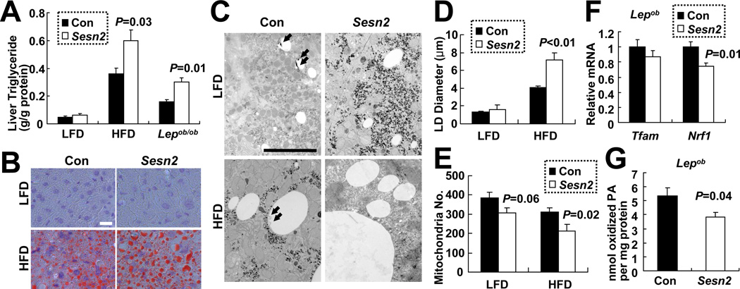
Figure 5. Sestrin2 attenuates hepatosteatosis during obesity.
(A) Elevated triglycerides in obese Sesn2−/− livers. Liver triglycerides in livers of Con (Sesn2+/+and Sesn2+/−) and Sesn2−/− mice kept on LFD or HFD, or Lepob/ob/Sesn2+/− or Lepob/ob/Sesn2−/− mice (n≥4). (B–D) Increased LD size in obese Sesn2−/− livers. (B and C) Liver LDs were visualized using Oil Red O staining (B) and EM (C) in Con and Sesn2−/− mice kept on LFD or HFD. Putative autophagosomes associated with LDs in Con livers are indicated with arrows. Scale bars, 20 µm (white), 5 µm (black). (D) Quantification of maximum LD diameters from EM images (21.4 × 31.9 µm, n≥8). (E and F) Reduced expression of mitochondrial biogenesis marker genes in obese Sesn2−/− livers. (E) Quantification of mitochondria number per field from EM images. (F) Relative Tfam and Nrf1 mRNA amounts in livers of indicated mice (n≥7). (G) β-oxidation of C14-labeled palmitic acid (PA) was measured in liver homogenates of Lepob/ob/Sesn2+/− (Con) and Lepob/ob/Sesn2−/− (Sesn2) mice (n≥5). Data are presented as means ± standard error. P values were calculated using Student’s t-test.

