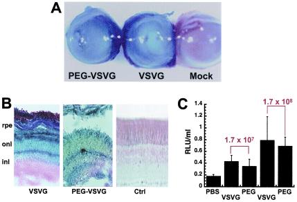
FIG. 3.
PEGylation does not compromise transduction efficiency of VSV-G-HIV in vivo. (A) Photomicrograph of whole eyes demonstrating grossly positive staining for beta-galactosidase expression from animals treated with PEGylated lentivirus (PEG-VSVG) and unmodified virus (VSVG) at a concentration of 1.7 × 10⁸ TU/ml and the absence of gene expression after injection of PBS (Mock; negative control). (magnification, ×100). (B) Histological sections indicate that transduced cells from eyes treated with native virus (VSVG) are located predominantly in the outer nuclear layer (onl), while those from eyes treated with the PEGylated virus (PEG-VSVG) are located throughout the retinal pigment epithelium (rpe), the outer nuclear later (onl), and the inner nuclear layer (inl) compared to sections from animals dosed with saline (Ctrl) (magnification, ×200). (C) Quantitative analysis of beta-galactosidase gene expression in eyes 7 days after treatment with either unmodified virus (VSVG), PEGylated virus (PEG) at two different doses (1.7 × 10⁷ and 1.7 × 10⁸ TU/ml), or PBS. The data are reported as the average number of RLU per milliliter of homogenate from five animals per treatment group and were normalized for protein content. The error bars represent the standard deviations of the data.

