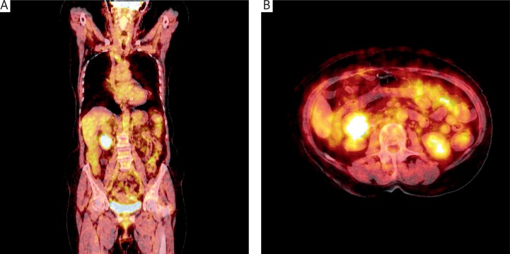
Fig. 1.
Coronal (A) positron emission tomography/computed tomography with the use of 18F-fluorodeoxyglucose (FDG PET/CT) images of the trunk and transverse (B) FDG PET/CT images of the abdominal cavity showing abnormal mass. Focal FDG uptake in the tumour (Nuclear Medicine Department, The Oncology Center, Bydgoszcz)
FDG – 18F-fluorodeoxyglucose; PET/CT – positron emission tomography/computed tomography

