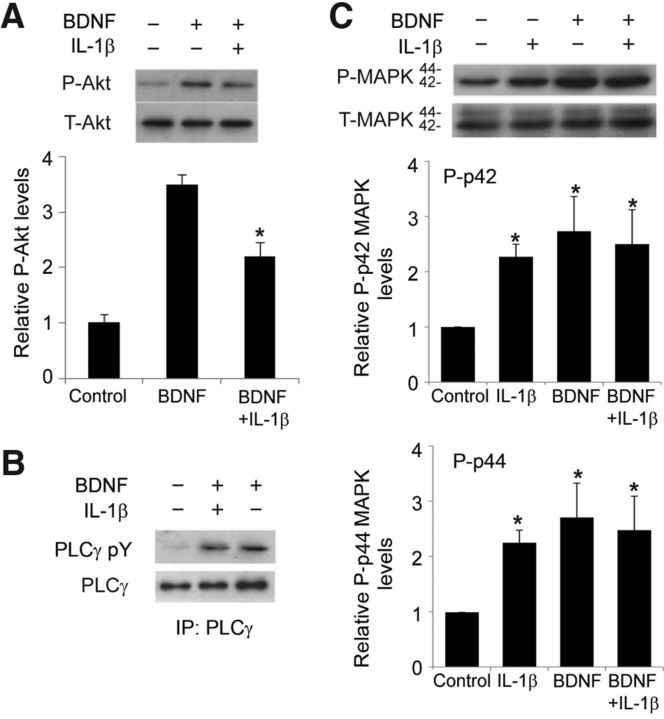
Figure 3.
IL-1β pretreatment affected BDNF-induced Akt activation. A, Representative Western blots showing the detection of P-Akt. Quantification of P-Akt levels showed that exposure to BDNF (100 ng/ml, 1 h) increased the amount of P-Akt, whereas pretreatment with IL-1β suppressed the effect of BDNF on P-Akt but had no effect on T-Akt. *p < 0.05 BDNF versus BDNF plus IL-1β (n = 4). B, Western blots showing that BDNF-induced (100 ng/ml, 1 h) tyrosine phosphorylation of PLCγ is unaffected by IL-1β pretreatment (50 ng/ml, 24 h) in hippocampal slices. Tyrosine phosphorylation of PLCγ was examined by immunoprecipitation (IP) followed by Western blotting, as described in Materials and Methods. C, Representative Western blots and quantification of the levels of P-p42 and P-p44 isoforms of MAPK in hippocampal slices treated as indicated. Data are the mean ± SEM (n = 3) expressed in terms of P-MAPK levels obtained in the control cultures. *p < 0.05 for all treatment groups versus control, ANOVA followed by the LSD post hoc test.

