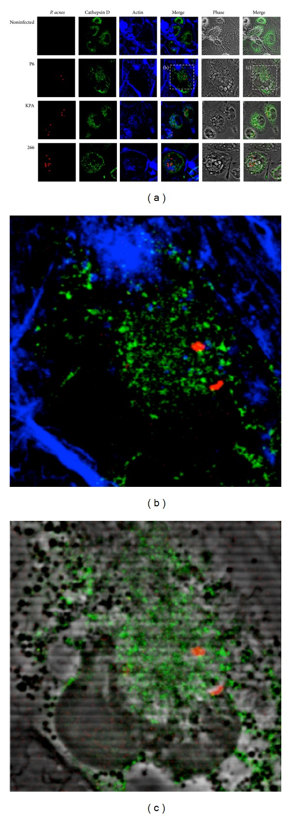
Figure 6.

Confocal analysis of P. acnes and cathepsin D in THP-1 cells at 24 h p.i. (a) Experiments were performed as described in Figure 5 legend. The lysosomal marker cathepsin D was stained with rabbit-anti-cathepsin D antibody and Cy2-conjugated donkey-anti-rabbit antibody (green). Intracellular P. acnes P6 (red), actin (blue). (b) and (c) Zoom-in on cells infected with P. acnes P6.
