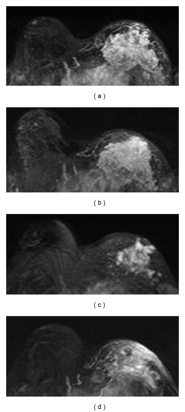
Figure 4.

A 29-year-old patient with non-mass-like enhancement lesion in the left breast. From (a) to (d), the maximum intensity projection (MIP) images of pretreatment, F/U-1, F/U-2, and F/U-3 MRI are shown. The extent of tumor size is 8.2 cm before treatment, remains about the same at 8.0 cm in F/U-1, shrinks down to 4.5 cm in F/U-2, and progresses again to 6.2 cm in F/U-3. The choline measured by MRS shows [tCho] = 0.77 ± 0.11 mmol/kg before treatment, which decreases to 0.20 mmol/kg in F/U-1, and then increases to 1.01 mmol/kg in F/U-2, and further increases to 1.70 mmol/kg in F/U-3. The transient decrease of tCho in F/U-1 precedes the size reduction observed later in F/U-2. And then the increase of tCho in F/U-2 indicates treatment failure, and the tumor grows larger in F/U-3.
