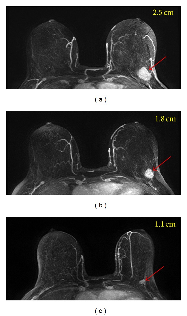
Figure 5.

A 64-year-old patient with a well-circumscribed mass lesion (invasive ductal cancer) in the left breast. From (a) to (c), the maximum intensity projection (MIP) images of pretreatment, F/U-1, and F/U-2 MRI are shown. The tumor size is 2.5 cm before treatment and shows concentric shrinkage to 1.8 cm in F/U-1 and further down to 1.1 cm in F/U-2 after completing treatment. The residual tumor size determined in post-NAC pathological examination is 1.4 cm. For mass lesion that shows concentric shrinkage, MRI is accurate in diagnosing residual disease.
