
Figure 2.
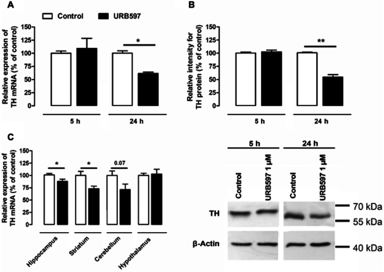
In vitro and in vivo URB597-mediated modification of TH expression. Quantifications of TH mRNA (A) and protein (B) expression were performed on neuroblastoma exposed for 5 or 24 h to 1 μM URB597. Quantitative PCR of TH mRNA was performed on total RNA extracts. The expression of TH mRNA (A) was normalized against GAPDH expression and results are given as the relative expression of treated versus control cells/animals. The densitometric analysis of the TH protein signals (60 kDa) shown in (B) was normalized against the measured signals corresponding to actin (42 kDa). A typical immunoblot is shown at the bottom right corner. Results are given as the percentages of expression relative to control cells. Data shown are means with SEM values of three experiments performed in triplicate. Two-tailed paired Student's t-test, *P < 0.05, **P < 0.01, relative to control at corresponding time (P = 0.0314, t = 5.507, residual d.f. = 2) and (P = 0.0087, t = 10.64, residual d.f. = 2) for mRNA and protein dosages respectively. In mice, TH mRNA contents were evaluated in hippocampus, striatum, cerebellum, cortex and hypothalamus tissues (C) 24 h after a single injection of URB597 (3 mg·kg−1, i.p.). Results are given as the percentages relative to control animals injected with vehicle only. Values are means with SEM of seven animals in each group. Two-tailed unpaired Student's t-test, *P < 0.05, relative to control (P = 0.0305, t = 2.452, residual d.f. = 12) (P = 0.0153, t = 2.824, residual d.f. = 12) (P = 0.0705, t = 1.985, residual d.f. = 2) in the hippocampus, the striatum and the cerebellum respectively.