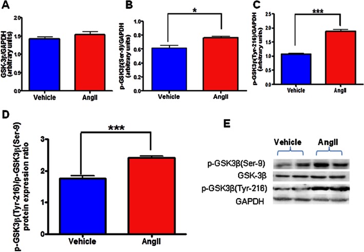
Figure 4.
Effects of AngII treatment on total and phosphorylated GSK-3β expression in neuronal cells. Serum-starved CATH.a cells were stimulated with 100 nM AngII for 6 h and cell extracts were then subjected to protein analysis by Western blot. Densitometric analysis of Western blot results showing protein expression of (A) total GSK-3β, (B) p-GSK3β(Ser-9), (C) p-GSK3β(Tyr-216), (D) p-GSK3β(Tyr-216)/p-GSK3β(Ser-9) ratio, and (E) a representative Western blot. AngII caused significant activation of GSK-3β as indicated by reduced p-GSK3β(Ser-9), increased p-GSK3β(Tyr-216), and increased ratio of p-GSK3β(Tyr-216) to p-GSK3β(Ser-9) protein expression in CATH.a neurons. AngII exposure did not alter total GSK-3β protein levels. The results are means ± SD of three independent experiments. *P < 0.05; ***P < 0.001 compared with cells treated with vehicle.

