
Fig. 1.
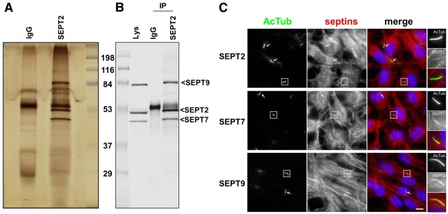
A SEPT2/SEPT7/SEPT9 complex at the primary cilium of RPE1 cells. (A,B) RPE1 cells expressing SEPT2–S-tag–GFP were extracted and proteins immunoprecipitated either with control antibody (IgG) or GFP followed by S-beads (IP), and proteins separated by SDS-PAGE and processed for silver staining (A), or western blotting with a mixture of antibodies to SEPT9_v1, SEPT7 and SEPT2 (B). (C) RPE1 cells, grown on coverslips and serum-starved for 24 hours, were processed for immunofluorescence using anti-acetylated tubulin antibody to stain cilia (AcTub, green), antibodies against SEPT2, SEPT7 or SEPT9 (red), and DAPI (blue) to stain the nuclei. Panels on the right are enlarged views of representative cilia (boxed in the main images). White arrows indicate other cilia in the same field. Scale bar: 5 µm.