Fig. 4.
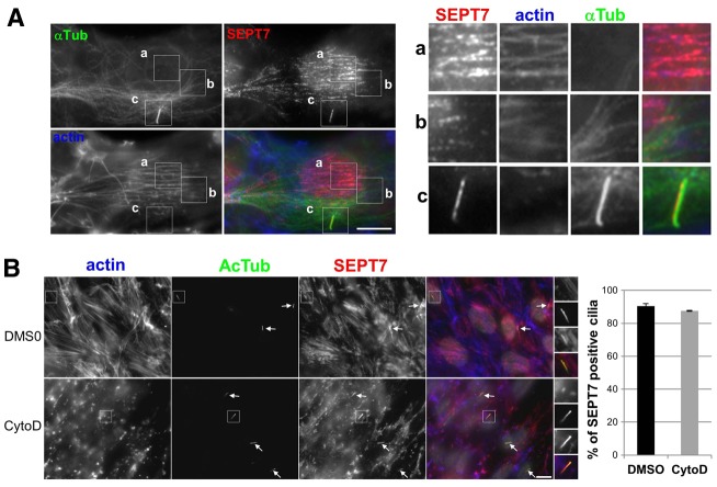
Localization of septins to the primary cilium does not depend on actin polymerization. (A) RPE1 cells were serum starved for 24 hours and then fixed and stained for immunofluorescence using antibodies against SEPT7 (red), α-tubulin (α-Tub; green), and fluorescent phalloidin to stain actin filaments (blue). Panels on the right are enlarged views of representative regions of the same cell (boxed in the main images) including stress fibers (a) microtubules (b) and the cilium (c). (B) RPE1 cells were serum-starved for 24 hours and then treated with 5 µM cytochalasin D (CytoD) or, as a control, with the same final concentration of DMSO, for 30 minutes at 37°C, then fixed and analyzed by immunofluorescence using antibodies against SEPT7 (red), acetylated tubulin (AcTub; green) and fluorescent phalloidin (actin; blue). Panels on the right are enlarged views of representative cilia (boxed in the main images). White arrows point to cilia in the same field. Scale bars: 5 µm. (Far right) Results were quantified (i.e. proportion of SEPT7-positive cilia) from three independent experiments (n = 40 cilia per experiments).

