Fig. 9.
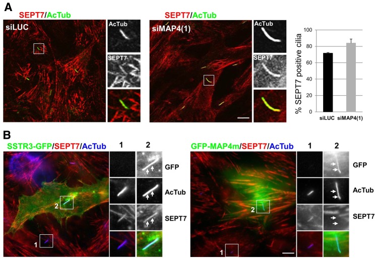
MAP4 competes with septins at the axoneme. (A) RPE1 cells treated with siMAP4(1) or luciferase targeting siRNA (siLUC) as a control (as in Fig. 8) were stained with antibodies against acetylated tubulin (AcTub, green) and SEPT7 (red). Panels on the right are enlarged views of representative cilia (boxed in the main images). The proportion of SEPT7-positive cilia was quantified in both conditions from three independent experiments (n = 30 in each experiment, P>0.1). (B) RPE1 cells were transiently transfected with SSTR3-GFP or GFP-MAP4m plasmids, fixed and processed for immunofluorescence with antibodies for SEPT7 (red) and acetylated tubulin (AcTub; blue), and the nuclei were stained with DAPI (blue). Panels on the right are enlarged views of representative cilia (boxed in the main images). White arrows indicate the position of cilia. Scale bars: 5 µm.

