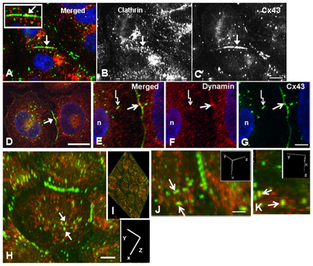
Fig. 1.
Immunocytochemical colocalization of the Cx43 protein with clathrin or dynamin in human adrenal tumor cells. (A–K) Colocalization of Cx43 protein (C and green in A) with clathrin (B and red in A) or dynamin (red in D–K) is shown. Note the colocalization (yellow) in the merged images with clathrin (A) and dynamin (D,E,H–K) in an area of the gap junction plaque (arrows in A–G) and annular gap junction vesicles (crooked arrows in E–G). Confocal microscopy techniques coupled with image rotation (a function within Nikon Elements that allowed us to reconstruct 3D images) shows the colocalization of dynamin at or near the equator of the annular gap junction vesicles (arrows in H,J,K). The axis of rotation is shown in the inserts in H–K. Scale bars: 10 µm (A–C,E–H); 4 µm (D); 5 µm (J,K).

